Screening workflow · Step 2 of 2
Analysis workspace
Screening SCR-DEMO-1046 · all visible results are prototype placeholders.
AI ENGINE NOT CONNECTED
Values shown in this prototype are interface examples only. No image analysis or medical prediction has been performed.
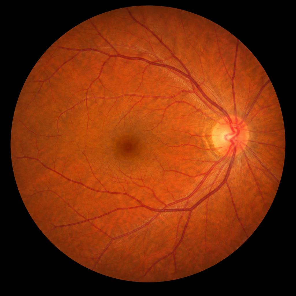
Original fundus image
Synthetic image for UI demonstration
DEMO DATA
100%
Screening summary
Prototype values only
Image qualityGood (demo)
DR severityModerate (demo)
Referable statusReferable (demo)
Confidence87% interface sample
Analysis statusDemo result only
Explainability
Future AI outputs will appear here
Grad-CAM
Attention map for model reasoning.
Lesion evidence
Lesion-level visual evidence.
Structure map
Retinal vessel and structure analysis.
Confidence
Calibrated confidence will be connected later.
Analysis views
Visual layers prepared for future AI integration
Original image
Enhanced image
Grad-CAM attention map
Lesion evidence overlay
Vessel / structure overlay